King Charles Is 10X More Inbred Than You (Here’s Why That Matters) SS
For those wondering why inbreeding is actually bad, not just weird, but genetically dangerous, here’s what nobody explained. You’ve probably heard that King Charles III is more inbred than the average person. That claim got shared everywhere, but nobody told you what it actually means inside his cells. What breaks? What fails? What happens to chromosomes, organs, and faces when generations of family trees fold back on themselves? So, let’s fix that.
Right now, walking around in your body, you carry somewhere between 100 and 400 potentially lethal genetic mutations. Don’t panic. This is normal. This is everyone. These mutations sit quietly in your DNA doing absolutely nothing harmful. And the reason is elegant. You have two copies of nearly every gene. One from mom, one from dad.
When one copy carries a dangerous mutation, but the other copy works fine, the working version compensates, produces enough functional protein, keeps everything running. Geneticists call these hidden mutations recessive because they recede into the background. They’re masked by their healthier partner.
Here’s where it gets interesting. The word carrier describes someone with one broken copy and one working copy of a gene. Carriers are healthy. They show no symptoms. They usually have no idea they’re carrying anything at all. The math only turns dangerous when two carriers of the exact same recessive mutation have children together.
Each parent passes one copy to the child. Which copy is basically a coin flip. When both coins come up mutant, the child inherits two broken copies of the gene. No backup, no working version to compensate. This state is called homozygosity. From the Greek homos meaning same, zygodos meaning yolked together. The child is now yolked to two identical broken genes and the disease emerges.
Take cystic fibrosis. One of the most common severe recessive disorders in people of European descent. One in 25 Caucasians carries a single mutated copy of the CFTR gene. Most never find out. But when two carriers have children, each pregnancy carries a one in4 chance of producing a child with two mutated copies.
That child’s lungs will fill with thick mucus. Their lifespan, even with modern treatment, is measured in decades rather than a full life. The mutation was always there, hiding across generations in healthy carriers waiting. But here’s the thing about rare mutations. The rarer a mutation, the less likely two unrelated people are to both carry it.
If only one person in 500 carries a particular mutation, the odds of two random strangers both carrying it and meeting and reproducing are basically zero. Inbreeding changes that calculation catastrophically. When relatives reproduce, they share recent ancestors and those ancestors mutations. A great great grandmother who carried a rare mutation passes that mutation down multiple family branches.
When those branches reunite through marriage, both partners may carry the same mutation inherited from the same source. Suddenly, extremely rare mutations have a direct path to homozygosity. Geneticists measure this risk using something called the consanguinity coefficient. The probability that any given gene in a person is homozygous because both parents pass down the same version from a shared ancestor.
An average person in an outbred population has a coefficient somewhere around 0.1%. Essentially nothing. First cousins who marry produce children with a coefficient of 6.25%. One in 16 gene positions carrying two identical copies from their shared grandparents. Research consistently identifies this threshold, first cousin marriage, as the point where measurable health effects start appearing across populations.
Studies of first cousin marriages in Pakistani communities in the UK found children were twice as likely to have congenital abnormalities compared to the general population. This is inbreeding depression, the technical term for the accumulated biological costs of reduced genetic diversity. King Charles III sits above that first cousin threshold.
His parents, Queen Elizabeth II and Prince Philip, were third cousins through Queen Victoria and also second cousins once removed through King Christian the 9th of Denmark. His coefficient runs approximately 10 to 13%. Not alarming by Hapsburg standards, but still roughly 10 times higher than the average person you’d pass on a London street.
That comparison matters because the British royal family sits at the mild end of a spectrum that stretches to genetic catastrophe. But first, a tutor king whose reproductive failures reshaped English history. Between 1509 and 1547, Henry VIII’s wives experienced at least 11 documented pregnancies. Only three children survived infancy.
Mary Elizabeth Edward. The pattern of loss follows a signature that modern medicine would recognize instantly, though it remains theory, not confirmed genetic fact. Catherine of Aragon endured the worst of it. Between 1510 and 1518, she experienced at least six pregnancies. a stillborn daughter in 1510.
A son who lived less than two months in 1511. Another son born dead in 1513. A third stillborn son in 1514. The surviving Princess Mary in 1516, a daughter who died within days in 1518. One healthy early child surrounded by repeated losses. Anne Berlin followed the same trajectory. A healthy first daughter, Elizabeth, in 1533, then a son still born in 1534.
A miscarriage in 1536. Jane Seymour produced a surviving son on her first pregnancy, Edward V 6th, in 1537 before dying of apparent postpartum complications. Do you see the pattern? First pregnancies often succeeded. Subsequent pregnancies often didn’t. In 2011, anthropologist Katrina Banks Whitley and bioarchchaeologist Kira Kramer proposed a theory that explains this with remarkable precision.
They suggested Henry VIII may have been kelp positive, a blood type present in roughly 9% of Caucasian populations. Here’s how kell incompatibility works. Kelp positive fathers and kel negative mothers face a specific reproductive problem. The first pregnancy typically succeeds because the mother hasn’t yet developed antibodies against the kel antigen.
But during birth, fetal blood cells enter the mother’s circulation and trigger an immune response. In subsequent pregnancies, the mother’s anti-kll antibodies cross the placenta and attack the fetal blood cells. The mother’s body rejects her own pregnancy. This theory explains why Catherine’s first pregnancy nearly succeeded. Why Mary survived despite being a middle pregnancy, possibly kell negative herself and not targeted by antibodies.
Why subsequent pregnancies consistently failed. Why Anne Bolin’s first child survived but the others didn’t. We don’t have Henry’s DNA. This remains hypothesis, not proof. But the pattern fits Kell incompatibility so precisely that serious scholars have paid attention. There’s more. The Kell gene region can in rare cases be associated with Mloud syndrome, a progressive neurological condition causing cognitive decline, emotional instability, chronic pain.
If Henry inherited a variant affecting both kell expression and neurological function, it might explain his transformation from the athletic, cultured prince of his youth to the paranoid, cruel tyrant of his final years. His chronic leg ulcer appearing after a jousting accident in 1536 and never healing could represent either a Mloud symptom or simply a wound complicated by his increasing obesity.
Henry’s own ancestry contributed to his genetic vulnerability. His parents Henry VIIIth and Elizabeth of York were third cousins, both descended from John of Gaunt. The tutor line was already operating within a restricted gene pool. The irony cuts deep. Henry executed two wives, divorced two others, all in pursuit of a male heir, and his genes, not his wives inadequacies, may have been driving the succession crisis that reshaped English history.
But Henry’s case is speculation awaiting proof that may never come. For documented peer-reviewed evidence of inbreeding destroying a royal line, we turn to the Hapsburgs. The Spanish Hapsburg dynasty ruled from 1516 to 1700. Their marriage strategy was deliberate, relentless to preserve territorial holdings across Spain, Austria, and the Holy Roman Empire.
The family married within itself generation after generation. Uncle niece marriages were common. Double first cousin marriages where both sets of grandparents were siblings occurred repeatedly. The family tree doesn’t branch outward like a normal pedigree. It loops back on itself. A 2009 study by Gonzalo Alvarez and colleagues published in Plo reconstructed the Hapsburg pedigree and calculated consanguinity coefficients for every member of the Spanish line.
The researchers traced over 3,000 individuals across 16 generations, documenting every marriage that tied the web tighter. The numbers tell a story of progressive collapse. Philip I who founded the Spanish line in 1504 had a coefficient close to zero. His parents were unrelated. His son Charles I who also ruled as Holy Roman Emperor Charles V rose to 0.
025 or 2.5%. Already concerning. Charles I married his first cousin Isabella of Portugal. Their son Philip II reached 0.115 11 12% nearly double the first cousin threshold. Philip II then married his niece Anna of Austria. She was the daughter of his sister Maria. Uncle and niece their son Philip III hit a coefficient of 0.
218 22%. and Philip III, he married his own first cousin, Margaret of Austria. Their son, Philip IVth, reached 0.233. Philip IVth then married Mariana of Austria. Mariana was simultaneously his niece. She was the daughter of his sister, Maria Anna. When Philip IV married his own sister’s daughter, the family tree completed another loop.
Their son Charles II born in 1661 achieved a consanguinity coefficient of 0.254 25.4% mathematically equivalent to being the child of a brother and sister. Though his pedigree contained no single such union, centuries of accumulated cousin marriages, uncle niece marriages, double first cousin marriages, all stacking generation after generation, produced the same genetic collapse through smaller increments.
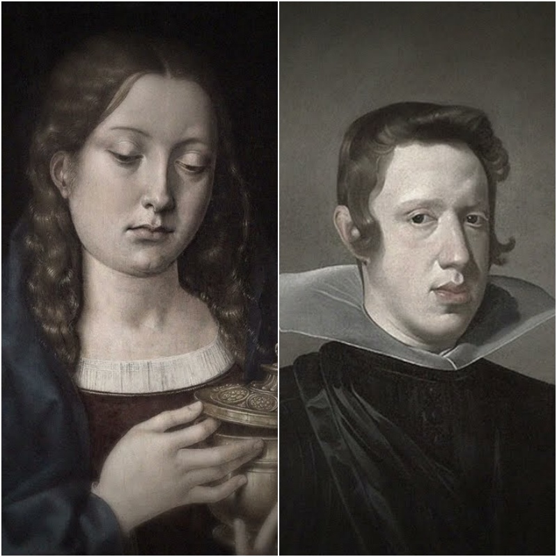
You can see the damage in every portrait. The Habsburg jaw, that dramatically protruding mandible that grew more pronounced across generations until Charles II could barely chew his own food. Mandibular prognism, the medical term, involves overgrowth of the lower jawbone relative to the upper jaw and skull.
The mandible extends forward and downward creating the characteristic underbite visible in every Habsburg portrait from Maxmillian I onward. A 2019 study led by Roman Velas at the University of Santiago de Compostella examined portraits of 15 Habsburg rulers alongside their calculated inbreeding coefficients. The correlation was unmistakable.
The more inbred the individual, the more severe the jaw deformity. But how does reduced genetic diversity actually deform bone? When many genes influence a trait, as with jaw structure, normal populations show a range of variations clustering around a functional average. Extreme outliers are rare because inheriting multiple unusual variants simultaneously is statistically unlikely.
Inbreeding changes those statistics. Homozygosity increases across the entire genome. Multiple unusual variants appearing together becomes far more probable. Developmental processes deviate from the functional average. Structures malform. Charles II’s condition extended far beyond his jaw. Born to Philip IV and Mariana of Austria, his own niece, Charles arrived with a constellation of problems.
He didn’t walk until age 8, couldn’t speak until age four. His enormous tongue made eating and speaking difficult throughout his life. Contemporary observers noted he appeared to have ricketetts, his legs buckling beneath him. He suffered chronic digestive problems that modern historians attribute to possible hormone deficiencies.

His father, Philip IV, had watched seven of his sons die before Charles was born. The dynasty’s ability to produce healthy heirs had been collapsing for generations. When Charles finally arrived, he was greeted not with celebration, but with cautious hope that this one might survive. He survived barely.
The immune system collapses at high inbreeding coefficients. Genetic diversity in the major hisystocompatibility complex, the gene region enabling immune recognition of pathogens, directly determines disease resistance. When inbreeding reduces MHC diversity, individuals become susceptible to infections that healthy immune systems easily control.
Studies of inbred populations consistently show higher rates of tuberculosis, pneumonia, and other infectious diseases. Charles was perpetually sick throughout his short life, constant illness, weakness, vulnerability to every passing contagion. This wasn’t bad luck. It was immune collapse written into his DNA.
Fertility fails through multiple mechanisms at high inbreeding coefficients. In males, sperm quality and quantity decline dramatically, reduced motility, increased rates of abnormal morphology. Studies show reduced sperm motility and the percentage of viable, properly shaped sperm falling. In females, miscarriage and still birth rates spike as embryos carrying lethal combinations of mutations fail to develop.
Charles married twice. first to Marie Louise of Orlon in 1679, then to Maria Anna of Newberg in 1689. Neither wife conceived. Doctors examined him. Priests prayed for him. Foreign powers watched anxiously, knowing the Spanish throne hung in the balance. His reproductive failure wasn’t chance. It was biological inevitability.
The heart and circulatory system accumulate defects in inbred populations at rates two to three times higher than outbred populations. Congenital heart defects, valve malf forations, septile abnormalities, holes between chambers that should be separate. The cardiovascular system, so complex in its development, offers countless opportunities for things to go wrong when genetic diversity disappears.
When Charles II died on November 1st, 1700 at just 38 years old, the autopsy painted a picture of systemic devastation. The physician reported finding a heart the size of a peppercorn, lungs corroded, intestines rotten and gangrous, three large stones in his kidney, a single blackened testicle, a head full of water.
Some of that description reflects 17th century medical understanding and the decomposition that had already begun. But the overall picture is consistent with failure across multiple organ systems. Exactly what population geneticists predict when inbreeding depression reaches severe levels. Charles II died childless.
The Spanish Hapsburg dynasty died with him. The War of the Spanish Succession followed, lasting 13 years and reshaping European politics for a generation. France, Austria, England, the Dutch Republic, all fought over the carcass of a kingdom whose ruling family had bred itself into extinction.
The dynasty that prioritized blood purity above all else had purified itself out of existence. But the Hapsburg collapse was visible, slow, documented across two centuries of narrowing gene pools and deforming jaws. Queen Victoria’s genetic legacy spread faster and its consequences reached further. Victoria reigned 63 years and produced nine children with Prince Albert, who was her first cousin.
Those children married into virtually every royal house in Europe, spreading British influence across the continent. They also spread a mutation in the gene coding for clotting factor 9, hemophilia B. Unlike the speculative kell theory for Henry, this mutation has been confirmed through DNA analysis of royal remains. Victoria was a carrier.
The gene sits on the X chromosome. Women have two X chromosomes, so a mutation on one can be compensated by the other. Men have only one X paired with a much smaller Y that carries different genes entirely. A single hemophilia mutation on a man’s only X chromosome leaves him without any backup copy, fully affected.
Victoria passed her mutant X to at least two daughters, Alice and Beatatrice, and to one son, Leopold. Leopold bled throughout his short life. Minor injuries became medical emergencies. A cut that would heal in days for others could bleed for weeks in Leupold. Joints swelled with internal bleeding after minor bumps.
He died at 30 from a brain hemorrhage after falling downstairs in 1884. A minor fall, a fatal outcome. Where did Victoria’s mutation come from? Her father, the Duke of Kent, showed no signs of hemophilia or carrier status. Her mother’s family had no documented history of the disease. Geneticists initially concluded it arose spontaneously, a denovo mutation during her father’s sperm production or her early embryionic development.
Recent research has suggested the alternative possibility that Victoria’s mother was herself a carrier who got lucky and never had an affected son. The question remains open. The consequences, however, are documented with precision that the Tutor and Hapsburg cases cannot match. The hemophilia spread through Victoria’s descendants like cracks through glass.
Alice passed the gene to her daughter Alexandra who married Zar Nicholas II and became Zarina of Russia. Beatatrice passed it to the Spanish and German royal lines through her children’s marriages. Within two generations of Victoria’s death in 1901, hemophilia had appeared in the ruling families of Russia, Spain, and Germany.
A single mutation from one woman fragmenting across a continent carried by the very intermarages that were supposed to strengthen royal bonds. The most famous case, Alexe Nikolavich Romanov, son and heir of Nicholas II and Alexandra, Victoria’s greatgrandson. Born August 12th, 1904. He inherited hemophilia from his mother, who had inherited carrier status from her mother, Alice, who had inherited it from Victoria.
Alexis’s condition would shape Russian history. The boy suffered his first serious bleeding episode at just 6 weeks old, bleeding from the navl for days after his umbilical cord fell away. By age two, a minor bump against a table left him with a massive hemorrhage in his leg. The internal bleeding created a hematoma so severe doctors feared amputation might be necessary.
He survived, but the pattern was set. Every bruise became a crisis. Every stumble a potential death sentence. Every childhood scrape that other boys forgot by dinnertime could send Alexi to bed for weeks. The Zarina became obsessed with protecting him. She restricted his activities, watched him constantly, lived in perpetual fear of the next bleeding episode.
The heir to the Russian throne could not play like other children, could not rough house with friends, could not learn to ride properly. The dynasty’s future hung by a thread of defective clotting factor. In 1912, while the family vacationed at their hunting lodge in Spala, Poland, Alexi jumped into a rowboat and injured his upper thigh against the gunnel.
Internal bleeding began and wouldn’t stop. For 11 days, the 8-year-old boy lay in agony as blood pulled in his tissues, creating pressure that crushed nerves and caused excruciating pain. His screams echoed through the lodge. Doctors could do nothing but watch. His mother, Alexandra, barely slept. She sat by his bed, holding his hand, watching him turn gray.
Court physicians prepared the family for his death. Official bulletins were drafted announcing the heir’s passing, held ready for release when the end came. Then Alexandra sent a telegram to Grigory Rasputin. Rasputin was a Siberian peasant who had cultivated a reputation as a faith healer and holy man. He had been introduced to the royal family in 1905 and had seemingly helped Alexi recover from previous episodes.
Whether through prayer, suggestion, or coincidence remained unclear. From his location hundreds of miles away, Rasputin responded by telegram. The illness is not as serious as it seems. Don’t let the doctors worry him. Within days, Alexi began recovering. Modern historians have speculated about what actually happened.
One leading theory, the court doctors had been administering aspirin for the boy’s pain. Aspirin thins the blood and inhibits clotting. Exactly the wrong treatment for a hemophiliac. Rasputin’s instruction to stop letting the doctors worry him, may have inadvertently stopped the aspirin treatment, allowing Alexis’s limited clotting ability to finally work.
Whatever the mechanism, Alexandra became convinced that Rasputin could save her son when medicine could not. The faith healer gained unprecedented access to the innermost circles of Russian power. His influence over the Zarina and through her over the Zsar became a source of scandal that eroded public confidence in the monarchy.
Rumors spread that Rasputin controlled the government, that Alexandra was his lover, that the holy manipulated state policy. None of it would have happened without hemophilia, a bleeding disorder caused by a mutation from a British queen creating political instability that contributed to the conditions for revolution.
And this brings us to a photograph that transforms abstract coefficients into visceral evidence. The 1913 wedding of Kaiser Wilhelm II’s daughter, Princess Victoria Louise in Berlin. In one image, King George V of Britain and Zar Nicholas II of Russia stand side by side in military regalia. They look like twins, not similar, not merely related, virtually identical.
Same beard trimmed to the same style, same eyes, same facial structure, same height, same bearing, same slightly melancholy expression. Contemporary observers who saw them together at family gatherings remarked they could barely tell the two men apart. During events where both were present, servants and guards reportedly confused them constantly.
This wasn’t coincidence. George and Nicholas were first cousins through their mothers Alexandra of Denmark and Dagmar of Denmark who took the name Maria Fodderona upon marrying into the Russian imperial family. The two Danish princesses were sisters, daughters of King Christian VI 9th. But the resemblance between George and Nicholas went deeper than mere cousinhood would explain in an outbred population.
Alexandra and Dagmar themselves looked nearly identical, and both George and Nicholas were also descended from Queen Victoria through additional lines. George was Victoria’s grandson through his father Edward IIIth. Nicholas had married Victoria’s granddaughter, Alexandra, a different Alexandra. Royal naming conventions recycled names endlessly.

The royal family trees of Europe had collapsed into a single extended family wearing different crowns. What the George Nicholas photograph demonstrates is the loss of phenotypic diversity, the visible variation in physical traits that healthy genetic mixing produces. In a genetically diverse population, first cousins can look completely different because they inherit different combinations of their grandparents thousands of genes.
The randomness of genetic recombination produces endless variety. When the gene pool shrinks, so does the range of faces. Christian the 9th of Denmark had so many descendants sitting on European thrones that he earned the nickname the father-in-law of Europe. His grandchildren ruled Britain, Russia, Greece, Norway, Denmark, and Spain.
And they all carried his genes, and Victoria’s jeans, mixing and remixing in ever tighter circles. By 1913, a family photo of European royalty looked less like an international gathering and more like a series of variations on the same face. 5 years after that photograph in Berlin, Nicholas was dead. Shot in a basement in Yakatarinburgg on July 17th, 1918 alongside his wife, his five children, and four servants.
The Bolevixs who ordered the execution feared the family might become a rallying point for counterrevolution. Alexe, the hemophiliac heir whose bleeding episodes had given Rasputin his power, was killed at age 13. The dynasty Victoria’s jeans had infiltrated through her granddaughter Alexandra, destroyed by revolution and bullets in a cellar.
George V survived. His line continues to the British throne today, which brings us back to King Charles III and his coefficient of approximately 10 to 13%. The Windsor inherited the narrow gene pool of Victorian era royalty. Elizabeth II and Prince Philip shared multiple common ancestors.
Queen Victoria, King Christian I 9th and others further back. Their marriage in 1947 continued the pattern of royal intermarriage that had characterized European dynasties for centuries. But something changed in 1981. Diana Spencer was marketed as a commoner bride. Though her aristocratic ancestry ran deep, she descended from Charles II through illegitimate lines and her family had been prominent in British nobility for centuries.
But she wasn’t closely related to Charles through recent ancestors. Her family tree connected to the royal line through different, more distant branches than the tight loops that connected Victoria’s great great grandchildren to each other. She brought genetic diversity the Windsor line had been lacking for generations.
and Catherine Middleton. Her family tree includes coal miners and factory workers alongside distant aristocratic connections. Her mother’s family were workingclass Londoners. Neither Diana nor Kate came from the closed loop of European royal intermarriage that produced the George Nicholas photograph. Prince William’s children, George, Charlotte, and Louie, carry significantly more genetic diversity than their greatgrandfather Charles’s generation.
The gene pool is opening again after centuries of narrowing. The pattern that destroyed the Hapsburgs and spread hemophilia across Europe has quietly been interrupted. Modern genetics has also transformed how consanguinius couples can approach reproduction. Carrier screening can now identify whether both partners carry the same recessive mutation before they have children.
Pre-implantation genetic diagnosis allows embryos created through IVF to be tested for specific genetic conditions before implantation. Couples who are closely related or who know they carry the same dangerous recessive mutation can use these technologies to have healthy children without the genetic lottery that destroyed Charles II.
The science that explained why the Hapsburgs collapsed now offers tools to prevent similar collapses. Genetic counseling helps families understand their risks. Prenatal testing can identify affected fetuses early in pregnancy. Gene therapy research aims to correct some of these mutations directly.
This isn’t about judging the royals. They didn’t understand genetics. Nobody did until the 20th century. What Victoria passed to her grandchildren, what the Hapsburgs accumulated across generations, what may have tormented Henry VIII’s marriages, none of it was intentional. They followed the political and social logic of their time which demanded that royal blood marry royal blood that territory be preserved through dynastic union that bloodlines remain pure.
They tracked those bloodlines obsessively while having no idea what those bloodlines were doing to bodies. But here’s what we can say now looking back with the tools of modern genetics. For roughly 500 years, European nobility conducted an unintentional genetic experiment. They married within their own small group generation after generation.
They watched jaws deform, children bleed, dynasties collapse, and attributed it to God’s will, to family curses, to the mysterious workings of fate. anything except the mathematics of shared DNA that we now understand. We have the documented results. Inbreeding coefficients aren’t trivia. They’re measurements of risk of how many positions in your genome might carry two identical copies of something that works better with variety.
The Habsburgs pushed past 25% and a dynasty died childless. The threshold for measurable problems starts around 6% first cousin level. The Windsor never approached Habsburg territory, but they accumulated enough shared ancestry across generations to make genetic diversity a genuine concern. One that appears to have been quietly addressed through marriages outside the traditional royal gene pool.
That’s what the math actually means. Not genealogy charts, not who married whom timelines. Chromosomes, organs, faces, the biological cost of keeping it in the family. Subscribe for more stories like